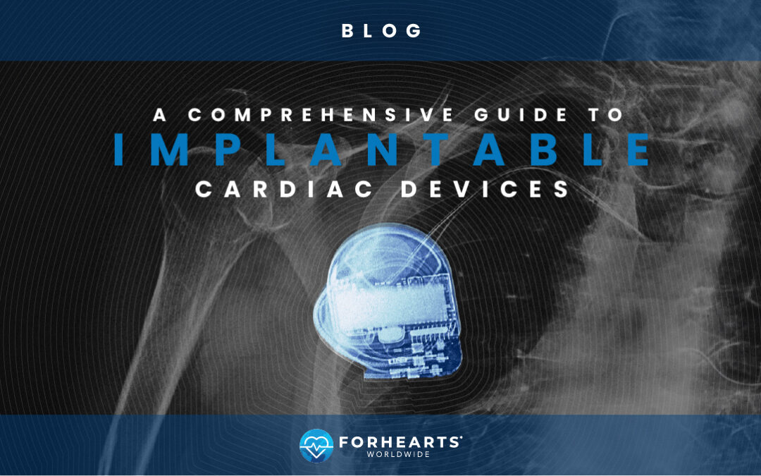
Millions of people worldwide suffer from heart issues that can severely impact and even shorten their lives. Fortunately, many heart conditions like arrhythmias, heart failure, coronary artery disease, and heart valve defects can be treated with implantable cardiac devices.
These sophisticated devices have revolutionized cardiac care, offering patients improved quality of life and longer lifespans. Let us learn more about the implantable cardiac devices that are saving lives.
Pacemakers
Implantable pacemakers regulate heart rhythm, ensuring a stable heart rate. Heart arrhythmia, where the heart has an abnormal rhythm, is the most common reason for a pacemaker. However, other cardiac issues can lead to irregular heart rhythm and slow heart rate (bradycardia). Heart transplant patients, as well as those suffering from an enlarged heart muscle, congestive heart failure, congenital heart defects, or have had a heart attack may have a weak heart that needs help to beat “in sync.”
Here is how pacemakers treat heart rhythm issues:
How Pacemakers Work
The heart is essentially an electrical system, with the sinus node, located in the right upper chamber of the heart, generating an electrical stimulus that contracts the heart (about 60 to 100 times a minute). Each contraction is one heartbeat. A pacemaker works by connecting to the heart’s electrical system via wires (called leads) and sending electrical pulses from the device’s pulse generator (housing the battery and the electronic circuitry) to electrodes inside the heart chamber, regulating the rate and rhythm of heartbeat. Pacemakers are not implanted into the heart but under the skin near the heart, usually at the collarbone.
Different Types of Pacemakers
Pacemakers are tailored to specific chambers of the heart, with the number of leads and contraction generation needed for a patient’s specific heart rhythm condition.
- Single-chamber pacemakers work on one chamber of the heart, with a single lead going to either the right atrium or the right ventricle.
- Dual-chamber pacemakers use two leads to connect to both the right atrium and the right ventricle, regulating both chambers of the heart. This helps the two chambers work together for more natural heart rhythms compared to single-chamber devices.
- Biventricular pacemakers synchronize the contractions of the left and right ventricles using three leads: one in the right atrium, one in the right ventricle, and one in the left ventricle. These pacemakers are most often used in patients suffering from heart failure.
- Rate-responsive pacemakers can adjust the pacing rate based on the patient’s activity level, ensuring that the heart rate increases during exercise and decreases during rest, mimicking the body’s natural response.
- Leadless pacemakers – These advanced devices are implanted directly into the heart without the need for external leads. They are designed to be less invasive and reduce complications associated with lead placement, making them a suitable option for certain patients.
Benefits of Pacemakers
Those suffering from irregular heartbeat often suffer from fatigue, dizziness, and fainting. In fact, these can be early symptoms of a heart rhythm disorder. Pacemakers help alleviate these symptoms and even improve the physical capabilities of patients. Besides improving quality of life, most importantly pacemakers extend lives. One study found that pacemakers restored life expectancy to normal levels for patients suffering from slow heart rhythm with no other heart issues or cardiovascular disease.
Implantable Defibrillators
While arrhythmia causes abnormal heart rhythm, dangerous arrhythmias like ventricular tachycardia and ventricular fibrillation can lead to sudden cardiac arrest if not treated. Part of that treatment can be an implantable cardioverter defibrillator, or ICD, which can shock the heart back into beating.
How Defibrillators Work
Defibrillators work and function like pacemakers, connecting to the heart’s electrical system via leads and sending electrical pulses from the device’s pulse generator to the heart. But ICDs can do more than regulate (or pace) an irregular heartbeat. These devices can deliver shocks to the heart, cardioverting it with a mild shock or defibrillating it with a stronger shock to restore normal rhythm.
Here’s how each function of an ICD works:
- Monitoring heart rhythm: The ICD continuously monitors the heart’s electrical activity through the leads, detecting various types of arrhythmias.
- Pacing: The ICD can act as a pacemaker, sending small electrical impulses to stimulate the heart to beat at a normal rate.
- Anti-Tachycardia Pacing (ATP): In patients with life-threatening fast heart rhythms, like ventricular tachycardia, the ICD can deliver a series of small, rapid pacing impulses to try to interrupt the arrhythmia and restore normal rhythm.
- Cardioversion: If ATP is unsuccessful or the arrhythmia is too fast and dangerous, the ICD can deliver a synchronized shock to the heart to restore a normal rhythm.
- Defibrillation: If the heart rhythm continues to be very fast and chaotic, the heart can go into ventricular fibrillation. The ICD can deliver a high-energy shock to the heart, resetting the heart’s electrical activity and restoring heart rhythm.
Different Types of Defibrillators
ICDs primarily treat arrhythmias that occur in the ventricles, the lower chambers of the heart. Like pacemakers, they can be wired to multiple chambers of the heart via multiple leads based on the need and severity of a patient’s heart rhythm condition.
- Single-chamber ICDs work on the lower chamber of the heart, with a single lead going to the right ventricle.
- Dual-chamber ICDs use two leads to connect to the right ventricle as well as the right atrium to treat both chambers of the heart.
- Biventricular ICDs provide cardiac resynchronization therapy to “resynchronize” contractions of the left and right ventricles and improve the heart’s pumping efficiency. These ICDs use three leads: one in the right atrium, one in the right ventricle, and one in the left ventricle. Like the biventricular pacemaker, these ICDs are most often used in patients suffering from heart failure.
- Subcutaneous ICDs are placed under the skin above the heart and attached to an electrode that runs along the breastbone. These devices are less invasive but can only deliver defibrillation shocks, as they are not connected to the heart’s electrical system via leads.
Benefits of Defibrillators
Patients suffering from dangerous arrhythmias due to heart failure or a weakened heart from heart blockages or heart attacks often have a poorer quality of life and shortened lifespan. ICDs can significantly improve survival rates for patients with these severe heart conditions. Most importantly, ICDs can prevent sudden cardiac death by immediately correcting life-threatening arrhythmias with a life-saving shock.
Ventricular Assist Devices
When the heart is weakened to the point that it can no longer regularly beat and pump blood sufficiently, a heart transplant may be the only option. Unfortunately, not everyone is a candidate for a heart transplant and those who are may spend years on a transplant list waiting for a heart. Ventricular assist devices (VADs) can serve as a stopgap until heart transplantation or even as an alternative to a heart transplant.
How Ventricular Assist Devices Work
Ventricular assist devices (VADs) are surgically implanted mechanical pumps that support the heart’s function of pumping blood throughout the body for those with weakened or failing hearts. Implanting and connecting a VAD to the heart requires open-heart surgery lasting several hours. Unlike pacemakers and defibrillators, VADs are not fully implantable, single-unit systems. To power the VAD, a computerized controller unit and battery pack are worn outside the body and connected back to the VAD via a drive line inserted through a small opening in the skin. The controller monitors the VAD’s function and provides low battery warnings.
How and where a VAD is connected to the heart depends on the severity of a patient’s heart failure.
Different Types of Ventricular Assist Devices
VADs can assist the left ventricle (LVAD), the right ventricle (RVAD), or both (BiVAD) to get the blood pumping and flowing to where it needs to go.
- Left Ventricular Assist Devices (LVADs) are the most frequently implanted and are placed in the left ventricle (the left lower heart chamber) to help pump blood to the aorta and, from there, to the rest of the body.
- Right Ventricular Assist Devices (RVADs) help the right ventricle in pumping blood into the pulmonary artery and then to the lungs. RVADs are usually temporary devices used during heart surgery or emergency care.
- Biventricular Assist Devices (BiVADs) use LVAD and RVAD in combination to help both the right and left ventricles move blood through the heart—pumping blood from the right ventricle into the lungs and the left ventricle out to the body. This is only used in severe cases of heart failure.
Benefits of Ventricular Assist Devices
By restoring or at least improving cardiac output and blood flow throughout the body, VADs can greatly enhance the quality of life for patients suffering from heart failure. This includes more energy, less fatigue, and better breathing and organ function. For those awaiting heart transplant, VADs are a crucial “bridge,” keeping patients alive and improving their quality of life while they wait for their new heart. For patients who are not eligible for a transplant, VADs can be a long-term option, with LVADs having a lab-tested device lifespan of 15 years and patients living with LVADs for as long as 13 years.
Artificial Heart Valves
As your heart is pumping, the heart’s four valves keep the blood flowing in and out of the heart’s chambers in the right direction. If a valve becomes narrowed, leaky, or stiffens and isn’t working sufficiently, the blood may not flow forward properly and instead flows backward into the heart chamber. When this happens, heart valve repair or even replacement surgery may be necessary.
How Heart Valves Work
The four heart valves are located between the heart’s four chambers:
- The tricuspid valve moves oxygen-poor blood flow from the right atrium in the heart’s upper chamber to the right ventricle in the lower chamber.
- The pulmonary valve ensures this oxygen-poor blood moves from the right ventricle to the pulmonary artery, which pumps blood to the lungs to get oxygen.
- The mitral valve ensures the now oxygen-rich blood coming back from the lungs moves from the left atrium in the upper chamber to the left ventricle in lower chamber.
- The aortic valve moves the oxygen-rich blood from the left ventricle to the aorta, which pumps blood out to the entire body.
Of these four heart valves, the mitral and aortic valves are the valves that are most often replaced. Replacing a heart valve requires open-heart surgery in which a cardiac surgeon removes the underperforming heart valve, replaces it with a new one, restarts the heart and ensures the replacement valve is working properly.
However, many heart valve patients are not good candidates for open-heart surgery due to age and/or weakened condition. The less invasive transcatheter aortic valve replacement (TAVR) or transcatheter aortic valve implantation (TAVI) procedures deliver a fully collapsible replacement valve through a catheter that is unfolded and sits atop the failed valve, seamlessly functioning in its place.
Different Types of Heart Valves
The type and size of a replacement valve depends on the heart valve that’s being replaced and the patient’s age and condition. Artificial heart valves can be either mechanical, biologically engineered, or come from donors.
- Mechanical valves are made from durable materials, usually carbon and metal, and last the life of the patient. However, they require recipients to be on lifelong anticoagulation therapy (blood thinners) to prevent blood clots. These valves are recommended for younger patients, as they lessen the chance of them needing another valve replacement, and for those already on blood thinners or with related cardiac or additional health issues that would make another heart valve procedure risky.
- Biological valves (also called tissue valves) are engineered from animal tissue and even animal valves. Because these valves are made from biological material, recipients do not require long-term blood thinning medication. However, they don’t have the lifespan of a mechanical valve, lasting roughly 10 to 20 years. For these reasons, tissue valves are considered best for older patients and those who can’t be on blood thinners.
- Donor valves come from human donors and last roughly 10 to 20 years, similar to biological valves. Human donor valves are often used for patients suffering from infections or conditions that affect the valve (rather than heart valve failure), replacing an infected valve with a healthy human one.
Benefits of Heart Valves
Replacing a failing heart valve can improve cardiac function and restore blood flow throughout the body, enhancing the quality of life for patients. This includes more energy, less fatigue, and better breathing, so replacement heart valve recipients are better able to perform the daily activities of life with improved overall health.
Stents
As the heart pumps blood out to the body, the blood runs through a series of passages—blood vessels that include arteries, veins, and capillaries. If these blood vessels become narrowed or weakened, the blood can’t circulate, and a blockage may occur. Inserting stents can keep these passages opened and blood flowing normally.
How Stents Work
A stent is a small, expandable tube that is inserted via a catheter through a sheath that is inserted into the narrowed blood vessel. X-ray is used to guide the catheter with the collapsed stent and deflated balloon to the blocked artery or blood vessel, where the balloon is inflated to widen the blood vessel and the stent is expanded and put into place to keep it opened. The stent will fuse with the body as tissue begins to coat it, and it will be fully lined within 3 – 12 months, depending on the type of stent.
Different Types of Stents
Stents can range in size and be made of different materials based on the part of the body and blood vessels they are being placed in.
- Fabric stents are used in larger arteries like the aorta.
- Silicone stents are often used in the lungs.
- Bare-metal stents are most often used for coronary arteries and made of metal mesh (like stainless steel, platinum-chromium, or cobalt-chromium) with no coating. These are the “original” stents, and the strong, resilient material nearly eliminates the risk of the artery collapsing.
- Drug-eluting stents are metal mesh stents that are coated with a polymer and medication that is slowly released to prevent scar tissue from growing around the stent and the artery possibly becoming blocked again. This improves long-term outcomes for the patient, making drug-eluting stents the preferred stents for coronary arteries.
Benefits of Stents
Blood vessel blockage can deprive the body’s tissues of vital oxygen-rich blood, leading to shortness of breath, fatigue, leg pain, chest pain, and, when the blockage is in the arteries, heart attack. By restoring proper blood flow, stents help alleviate these symptoms and reduce the risks of heart blockages and heart attacks.
Implantable Cardiac Devices For All
Implantable cardiac devices save lives. But too often, these medical technologies can be slow to make it to people and countries with limited resources. At ForHearts Worldwide, we are committed to the belief that no person should die because they do not have access to modern medical technology. For 40 years, we have been providing implantable cardiovascular devices for people in need in developing countries around the world.
Our mission is in our name—to do all we can ForHearts in need. Now, we are excited to announce our new partnership with the Pacemaker Club, an online community where people living with implantable cardiovascular devices can connect, share information, and support one other.
You can be part of the ForHearts mission supporting hearts in need around the world. Explore our donation opportunities.

